Aortic Valve Anatomy
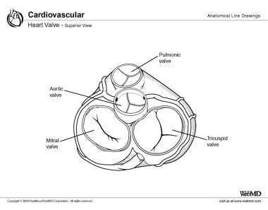
The normal human heart contains 4 valves that regulate blood flow into and out of the heart. The aortic and pulmonic valves are known as the semilunar valves, whereas the tricuspid and mitral valves are referred to as the atrioventricular valves. All the valves are trileaflet, with the exception of the mitral valve, which has 2 leaflets. Cardiac valves are surrounded by fibrous tissue forming partial or complete valvular rings, or annuli. These annuli join the fibrous skeleton of the heart to anchor and support the valvular structures.
The aortic valve is located between the left ventricular outflow tract and the ascending aorta. The aortic valve is the cardiac centerpiece. Relative to the aorta, the mitral valve is located posterior and to the left, the tricuspid valve is located inferiorly and to the right, and both valves abut on the posteroinferior margins of the aortic root, albeit with the atrioventricular separating structures interposing between the root and the tricuspid valve
In most cases, the orifices of the coronary arteries arise within the 2 anterior sinuses of Valsalva, usually positioned just below the sinotubular junction. However, arteries can occasionally be positioned superior relative to the sinotubular junction. As a result of the semilunar attachment of the aortic valvar leaflets, 3 triangular extensions of the left ventricular outflow tract reach the level of the sinotubular junction. These triangles are formed of thinned fibrous walls of the aorta between the expanded sinuses of Valsalva. Their most apical regions represent areas of potential communication with the pericardial space and, in the case of the triangle between the right and left coronary aortic leaflets, with the plane of tissue interposed between the aorta and anteriorly located sleeve-like subpulmonary infundibulum. The 2 interleaflet triangles bordering the noncoronary leaflet are also in fibrous continuity with the fibrous trigones, the mitral valve, and the membranous septum.

Embryology
Semilunar valve formation begins during the fourth week of gestation. At this time, opposing dextrosuperior and sinistroinferior endocardial cushions appear in the cephalad portion of the truncus arteriosus. Simultaneously, 2 additional intercalated endocardial cushions form, each located 90º from the aforementioned dextrosuperior and sinistroinferior endocardial cushions.
The dextrosuperior and sinistroinferior cushions fuse and, in doing so, form the truncal septum. The truncal septum undergoes a complex process of differentiation, eventually forming the right and left aortic valve cusps and 2 leaflets of the pulmonic valve. Of the 2 intercalated endocardial cushions, the right cushion eventually forms the posterior aortic valve cusp, whereas the left forms the anterior pulmonic valve leaflet. This occurs during the counterclockwise rotation and caudal shift of the conotruncus. During this time, the endocardial cushions also undergo dedifferentiation from a myosin-heavy chain to an alpha-smooth muscle actin phenotype, resulting in mature arterial valvular leaflets. The improper fusion or the incomplete dedifferentiation of the previously mentioned endocardial cushions is thought to be responsible for the formation of anatomically and structurally congenitally abnormal aortic valves.
The following image is an overview of the transitions occurring in early heart development in amniotes Within the right atrium, the atrioventricular node is located within the triangle of Koch. This important triangle is demarcated by the tendon of Todaro, the attachment of the septal leaflet of the tricuspid valve, and the orifice of the coronary sinus. The apex of this triangle is occupied by the atrioventricular component of the membranous septum. The atrioventricular node is located just inferior to the apex of the triangle adjacent to the membranous septum; therefore, the atrioventricular node is in close proximity to the subaortic region and membranous septum of the left ventricular outflow tract. This relationship explains the risk of developing complete heart block or conduction abnormality in patients who suffer from various pathologies involving the aortic valve. The atrioventricular node continues as the bundle of His, piercing the membranous septum and penetrating to the left through the central fibrous body, which runs superficially along the crest of the ventricular septum, giving rise to the fascicles of the left bundle branch.
The triangle between the right and noncoronary sinus is in close proximity to the bundle of His as it courses through the central fibrous body just below the inferior margin of the membranous ventricular septum, which can be of clinical significance in patients with endocarditis.
for more info


No comments:
Post a Comment